


Medical Equipment
As the leading hospital in cancer care, the 500-bed-capacity NCC Hospital provides the general public with the highest quality of care. Greater effectiveness in therapy and improved patient quality-of-life can be attributed to, among others, the state-of-the-art equipment. The hospital offers IMRT(Intensity Modulated Radiation Therapy), Tomotherapy, PET/CT, CT simulation, Gamma cameras, Coincidence Cameras and Superconducting MRI. Moreover, the NCC introduced the proton therapy system with a cyclotron capable of generating a 230-MeV proton beam at its Proton Therapy Center in march 2007. As the first and only establishment in Korea equipped with this most advanced radiation therapy facility, the NCC hospital will undoubtedly be able to better serve the various needs of cancer patients in Korea.

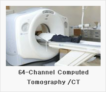
Tomotherapy
Tomotherapy looks like a CT scanner because it is a MV-CT scanner, allowing efficient 3D CT imaging to be used for ensuring the accuracy of treatment for every patient, every day. This is what is called the *Image-Guided Radiation Therapy (IGRT). IGRT allows doctors to better target the cancer while avoiding nearby healthy tissue. For this unique designed gantry, we can deliver the intensity-modulated radiation Therapy (IMRT) from all angles around the patient. Tomotherapy was designed so that highly-precise treatment plans could result in more effective treatment deliveries, with minimized side effects for patients.
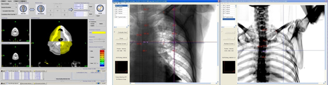
What is IGRT (Image Guided Radiation Therapy) technology
IGRT assists in delivering radiation therapy to cancerous tumors better than before. This is very useful since tumors can move between treatments due to differences in organ filling or movements while breathing and daily setup error. IGRT involves conformal radiation treatment guided by specialized imaging tests, such as CT scans, MVCT scans, ultrasound or X-rays. These tests are done in a treatment room just before a patient receives daily radiation therapy treatment. We can support the IGRT technology by using MVCT image in Tomotherapy. Also, Proton machine have orthogonal two X-ray imagers for precise cancer targeting.
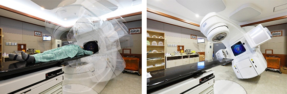
Linear Accelerators (Vital, Trilogy, True-Beam)
We also have 4 linear accelerators. This machine can make make high& low energy high & low energy X-ray (photon) and electron for cancer treatment. Additionally, 21EX Clinac can deliver Intensity-Modulated Radiation Therapy (IMRT). IMRT technique is the last trend in the world. It can modulate the radiation intensity at the tumor shape. The modulation increases cure rate with minimized side effects for patients.
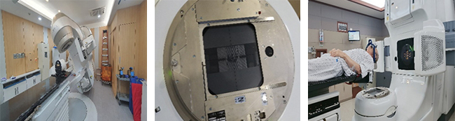
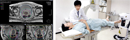
How to make beam-shapes for each patient
(Multi-leaf collimators systems and 3-Dimentional conformal technology ) (Intensity Modulated Radiation Therapy) In principle, every cancerous tumor has its own shape. Before the reatment, we use computers and special imaging techniques such as CT, MR or PET scans to show the size, shape and location of the tumor as well as surrounding organs.
Oncologist can then precisely tailor the radiation beams to the size and shape of your tumor with multi-leaf collimators.
We can provide 3mm, 5mm, 10mm sized multi-leaf collimators(MLC). Specially, 3mm sized (micro-size) MLC is dedicated for stereotactic radio surgery.
Intensity modulated radiation technique (IMRT) can modulate beam intensity during irradiation. It helps to save healthy tissue and give enough dose to tumor. As a result, we expect IMRT has much higher curable rate than 3D conformal radiation therapy.
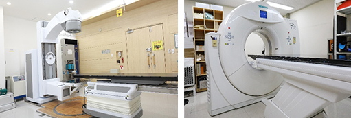
Conventional Simulator AND CT- simulator
We also have the 4 linear accelerators. This machine can make the high & low energy X-ray (photon) and electron for cancer treatment. Additionally, 21EX Clinac can delivers Intensity-Modulated Radiation Therapy (IMRT). IMRT technique is the last trend in the world. It can modulate the radiation intensity at the tumor shape. So increase the cure rate with minimized side effects for patients.
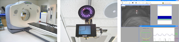
How to deliver beam to the target precisely in moving organs
(4D CT and Real-time Position Management Systems )
Breathing causes some organs keep going move during a treatment (ex. Lung, Upper abdominal Sites). In turn, we trace patient’s respiration cycles and make the treatment plan by reflecting that data in some cases.
4D-CT and Real-time Position Management Systems (RPM) allows clinicians to correlate tumor position in relation to the patient's respiratory cycle.
4D-CT provides clean images for planning so that clinicians can visualize the target more clearly with fewer of the image artifacts associated with respiratory motion.
This system also helps to minimize dose to healthy tissue during the radiation therapy. In addition, we have GE high speed 4-D CT scanner and Varian RPM systems that are widely used in the world.
What is the Brachytherapy?
Brachytherapy (from the Greek word brachy, meaning "short-distance"), also known as internal radiotherapy or sealed source radiotherapy, is a form of radiotherapy where a radiation source is placed inside or next to the area requiring treatment.
Brachytherapy is commonly used as an effective treatment for cervical, prostate, breast, and skin cancer and can also be used to treat tumors in many other body sites.
A key feature of brachytherapy is that the irradiation only affects a very localized area around the radiation sources. We have high-dose rate brachytherapy machine from Neucletron. It is armed with high-dose rate radiation source (Ir-192) which allows patient to be treated as outpatient- based.
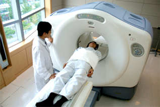
Positron Emissing Tomograph Computed Tomography (PET-CT)
PET-CT is a medical imaging device which combines in a single gantry system both a Positron Emission Tomography (PET) and an x-ray Computed Tomography, so that images acquired from both devices can be taken sequentially, in the same session from the patient and combined into a single superposed (co-registered) image. Thus, functional imaging obtained by PET, which depicts the spatial distribution of metabolic or biochemical activity in the body can be more precisely aligned or correlated with anatomic imaging obtained by CT scanning. Two- and three- dimensional image reconstruction may be rendered as a function of a common software and control system.
Magnetic Resonance Imaging (MRI)
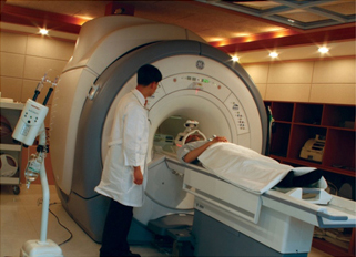
MRI is primarily a medical imaging technique most commonly used in radiology to visualize detailed internal structure and limited function of the body. MRI provides much greater contrast between the different soft tissues of the body than computed tomography (CT) does, making it especially useful in neurological (brain), musculoskeletal, cardiovascular, and oncological (cancer) imaging. Unlike CT, it uses no ionizing radiation, but uses a powerful magnetic field to align the nuclear magnetization of (usually) hydrogen atoms in water in the body. Radio frequency (RF) fields are used to systematically alter the alignment of this magnetization, causing the hydrogen nuclei to produce a rotating magnetic field detectable by the scanner. This signal can be manipulated by additional magnetic fields to build up enough information to construct an image of the body.
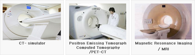
64-Channel Computed Tomography (CT)
The 64-Channel CT embodies completely the philosophies of sense and simplicity applied to more effective patient care. With a range of new and targeted dvancements, including low-dose cardiac imaging, 3D volume software and expanded portal capabilities, this system takes the raw capabilities of volume imaging and makes them work harder for patients by making the physician’s work easier. This system can expand clinical boundaries in cardiac, pulmonary, trauma, and pediatric imaging.